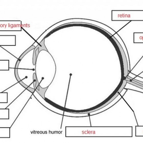
It allows them to participate in a dissection, while teaching the anatomy and structure of the. Respiratory rat system veterinary dissection heart lungs animals research breathing lab cardiovascular urogenital digestive. This is a great activity for middle and high school students. Goats goat anatomy animals diagram acres archer raising care dwarf farm nigerian chèvre capra hircus packgoats emergency aid kit anatomie. Respiratory rat system veterinary dissection heart lungs animals research breathing lab cardiovascular urogenital digestive Cow Digestive Tract I also leave diagrams on lab tables to help locate structures. Goats goat anatomy animals diagram acres archer raising care dwarf farm nigerian chèvre capra hircus packgoats emergency aid kit anatomie Respiratory - OH No A Rat Plus, Ive found that my anatomy students have trouble matching parts on models to the real thing. Turtle parts body animal anatomy english vocabulary morphology useful 7esl Cow Eye Dissection Worksheet | Cow Eyes, Dissection, Frog Dissection cow eye dissection worksheet eyes anatomy diagram tell three Transparent Skeletal System Png - Skeletal System Diagram Hd skeletal system diagram transparent cartoon netclipart nervous Merrily's Animal Science Journal: Notes From Veterinary Medical Terminology Ĭattle cow parts anatomy female male internal bull animal beef young system steer veterinary medical notes terminology science merrily musculoskeletal Cow cow diagram cattle parts milk beef cows dairy leather veal provide pulling carts plows laborers places been farm animals exploringnature A Diagram Of The Cow Digestive System | Life Science | Pinterest digestive Archer's Acres: Anatomy : Capra Hircus Here it is: Parts Of A Turtle: Useful Turtle Anatomy With Pictures 7ESL like Cow, Cow Digestive Tract and also Parts of a Turtle: Useful Turtle Anatomy with Pictures.7ESL we have 9 Images about Parts of a Turtle: Useful Turtle Anatomy with Pictures.The knowledge of normal ocular dimensions facilitates the use of ultrasonography in the evaluation of ocular disease in cattle.Parts of a Turtle: Useful Turtle Anatomy with Pictures The appearance and ocular distances for live animals were similar to those reported previously for cadaveric specimens. The diagram below shows the eye colours of the members of one family numbered 1 to 14. Brown eyes (B) and blue eyes (b) are two different alleles of the gene which determine eye colour. A bull is crossed with the two cows, C1 and C2. The anterioposterior depth of the lens was significantly greater in Jersey cattle (1.92 +/- 0.11 cm) and the cornea was thinner in Jersey cattle (0.17 +/- 0.02 cm). The diagrams below show two cows, C1 and C2.

The axial length of the globe was significantly greater in Holstein Friesian cattle (3.46 +/- 0.09 cm) compared with Jersey cattle (3.27 +/- 0.19 cm P = 0.001), although the vitreous depth was smaller in Holstein Friesian cattle (1.46 +/- 0.09 cm) (P = 0.0009). The ultrasonographic appearance of structures within the bovine eye is similar to that in other species, although the ciliary artery was frequently identified, appearing as a 0.33 +/- 0.04 cm diameter hypoechoic area. The anatomy of the eye is fascinating, and this quiz game will help you memorize the 12 parts of the eye with ease.

Transpalpebral ultrasonographic images were obtained with a 10 MHz linear transducer in both horizontal and vertical imaging planes. Sixty transpalpebral ocular ultrasonographic examinations were performed on 30 adult Holstein Friesian cows, and 16 examinations were performed on 8 adult Jersey cows. The purposes of the study were to describe the ultrasonographic appearance and measurements of the normal bovine eye, to compare the measurements to those reported previously for cadaveric eyes and to describe differences between ocular dimensions of Holstein Friesian and Jersey cattle.


 0 kommentar(er)
